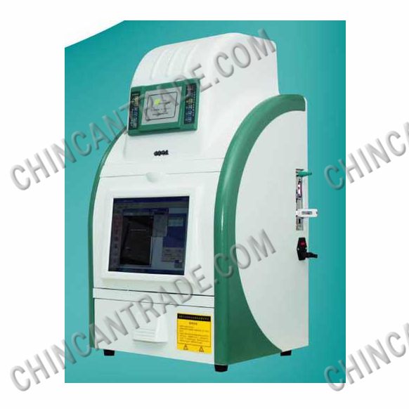
JS-3000
Automatic Gel Imaging Analysis System

JS series’ gel imaging analysis system acquires various nucleic acid and albumen electrophoresis images directly through JS gel imaging analyzer. System configures advanced digital CCD to assure the sensitivity of gel images taken under low-light. System configuration of 12 inch completely touching display screen makes observation of gelling very convenient without external computer. Embedded with industrial computer achieves completely integration designing, we need not to buy another computer and can truly realize the integration of shooting, storing and analyzing. This machine can be placed in narrow space, installing it very convenient and fast, just need take gel imaging Analyzer out from package, access power and turn on the switch, then it can be used. Needn’t the help of installing, needn’t training, let alone engineers’ service, users can easily master how to use this product. All the processes of shooting, storing and analyzing controlled through touching display screen, it can connect to printer, exchange various images and data files through USB and network. Application of advanced network teaching and distance diagnosis technology makes you no anxiety forever.
| JS-3000 Automatic Gel Imaging Analysis System |
| Name & Specification |
| JS-3000 Automatic Gel Imaging Device |
| SONY ICXCCD professional digital CCD |
| Data-processing system |
| Computer M6Z1212M 2/3 inches, large diameter, high permeability of lens, F=1:1.2, 12.5-75mm |
|
Coating filter specially designed for various fluorescent dye of gel imaging feature , 1 bit light filter wheel(optional of manual 5 bits light filter wheel and automatic 5 bits light filter wheel controlled by computer) |
| Gel cutting devices |
| Gel Image Shooting Software |
| Gel Imaging Analysis Software |
Technical parameters
| JS-3000 Automatic Gel Imaging Analysis System | |
| Digital CCD | SONY ICXCCD professional digital CCD |
| Detection Sensitivity | The detection of Nucleic acid as Low as 0.02ng |
| Effective Pixel | 1392 x 1040 |
| Data-processing system | Intel Atom N270 1.60GHz,512kbyte L2 |
| Memory | 1 x DDR2 SODIMM 1GB (Maximum capacity 2GB) |
| Hard disk | Solid hard disk 8GB(or Solid hard disk 120GB) |
| USB | Support seven USB 2.0(three USB 2.0 need be expanded) |
| NIC | 8111C Realtek on-board double -gigabit NIC, support no disk |
| Pixel size | 4.6μm x 4.6μm |
| SNR | ≥ 60dB |
| Shutter control | Electronic shutter |
| Exposure time |
Long exposure time from 4ms to 60 minutes, collect and store all images successively, possess image correction function |
| Interface | Standard C interface |
| Cutting device | special observation and cutting devices (visual angle and the object is 90 °) |
| Color filter film |
Coating filter specially designed for various fluorescent dye of gel imaging feature ,Standard configuration of 590nm(or 535nm, 620nm, 460nm) EB/SYBR Green, BP |
| Light filter location |
1 bit light filter wheel (optional of manual 5 bits light filter wheel and automatic 5 bits light filter wheel controlled by computer) |
| Case control |
Via case panel to realize the automatic control - zoom, focus, aperture, transmission UV lamp and reflector lamp |
| Computer control |
highly programmed (computer controlled black box / power / UV and white light switch / iris / focus / focal length) |
| Timing shutdown |
Shutdown timer for 15 minutes, effectively extending the UV lamp life and UV glass |
| The way of Opening door | pull-down to open the door and drawer-platform |
| UV transmission loading board |
Transmission wavelength: 302nm(254nm, 365nm (is optional)), size: 20cm x 25cm(special specifications can be customized) |
| White light reflection | Cooling light, voltage 12V |
|
White light transmission loading-board |
Cooling light, voltage 12V, chassis panel internal folding 20cm x 25cm (special specifications can be customized) |
| Quality authentication |
Only one of gel imaging systems passes national authoritative measurement authentication instruction, and obtains third class of Shanghai scientific advance prize |
| Shape size | L50CM X W37CM X H82CM |
| Application |
can be used for DNA / RNA gels, protein gels, blot autoradiography film plastic card, the enzyme Standard plate, thin layer chromatography plate, imaging and analysis of dish |


